Who it is for
The two forms of the condition require different approaches:
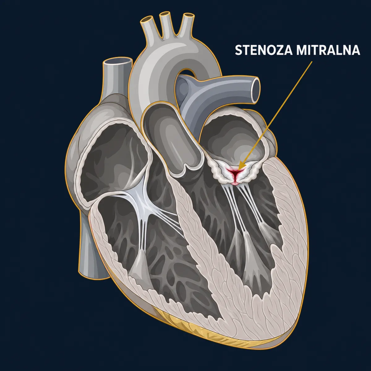
- Rheumatic — the leaflets are fused at the commissures (the points where they meet), thickened, with involvement of the chordae tendineae; it mainly affects middle-aged people, more often women. It is suitable for separating the commissures.
- Degenerative — massive mitral annular calcification (MAC) in older people, often with coronary artery disease and kidney failure; the commissures are not fused, so commissurotomy does not help, and surgery is technically difficult.
Procedural treatment is considered for patients with significant stenosis (valve area ≤ 1.5 cm²) who:
- have symptoms — breathlessness on exertion, fatigue, palpitations, coughing up blood;
- have no symptoms but are at high risk of complications: new atrial fibrillation, a previous embolic event, pulmonary hypertension, a planned pregnancy or major non-cardiac surgery.
What it involves
- Percutaneous mitral commissurotomy (PMC, balloon valvuloplasty) — the first-choice method in rheumatic disease when the valve structure is favourable (flexible leaflets, little calcification, no significant regurgitation, no clot in the left atrium). A balloon is introduced through the femoral vein and a puncture in the wall between the atria, and it splits the fused commissures. The procedure is performed by interventional cardiologists.
- Surgery — when the valve is not suitable for commissurotomy, when there is coexisting significant regurgitation, a clot in the atrium, or a need for another heart operation. Most often valve replacement (a mechanical or biological prosthesis, preserving the subvalvular apparatus); surgical commissurotomy (open, under direct vision) is rarely performed today. In degenerative stenosis with massive annular calcification, surgery requires special techniques, and in high-risk patients transcatheter implantation of a valve into the calcified annulus is considered (an evolving method, in reference centres).
- During surgery, atrial fibrillation ablation and left atrial appendage closure are usually performed.
Surgical access: sternotomy or right mini-thoracotomy, as in mitral regurgitation.
What happens
Percutaneous commissurotomy: local anaesthesia with sedation, 1–2 hours, guided by transoesophageal echocardiography; beforehand, a clot in the left atrial appendage must be ruled out. Hospital stay 1–3 days.
Surgery: admission the day before; tests as before any valve operation (transoesophageal echocardiography, coronary angiography, CT angiography for minimally invasive access). General anaesthesia, cardiopulmonary bypass, 3–4 hours. One day in intensive care. Hospital stay 6–8 days.
Recovery
- After commissurotomy: return to activity within a few days; echocardiographic follow-up after the procedure and every year — the stenosis may recur after years, and the procedure can then be repeated.
- After surgery: protect the breastbone for 6–8 weeks (after sternotomy); cardiac rehabilitation.
- Anticoagulation: in mitral stenosis with atrial fibrillation — lifelong, and with a vitamin K antagonist (warfarin/acenocoumarol), because NOACs (non-vitamin K oral anticoagulants) are not recommended in moderate or severe rheumatic stenosis or with a mechanical prosthesis. After a biological prosthesis without atrial fibrillation — usually 3 months. The regimen is set by your doctor.
- Prevention of endocarditis; in patients who have had rheumatic fever — prevention of recurrence, as recommended.
Risks and alternatives
Percutaneous commissurotomy: success 80–95% with good anatomy; complications — significant regurgitation requiring surgery 2–10%, tamponade, embolism (< 1–2%), a persistent small hole in the atrial wall (usually of no significance).
Mitral valve replacement surgery: perioperative death 2–5% depending on the patient's condition and pulmonary hypertension; stroke 1–3%; bleeding requiring re-operation 3–5%; pacemaker 3–5%. In degenerative stenosis with massive calcification, the risk is clearly higher.
Alternatives: conservative treatment — diuretics, rhythm control, anticoagulation — relieves symptoms but does not remove the stenosis; appropriate for mild disease or when a procedure is not possible.
More on how the choice between surgery and a transcatheter procedure is made →
Questions
Why can't I take the "new" anticoagulants with this condition?
Because the studies that showed NOACs to be safe in atrial fibrillation excluded patients with moderate and severe rheumatic mitral stenosis and those with a mechanical prosthesis. In these groups, warfarin or acenocoumarol with INR monitoring remains the standard.
Does commissurotomy last a lifetime?
Usually not; it gives a good result for 10–15 years and can be repeated. Valve replacement is a definitive solution, but it comes with the burden of a prosthesis.
Does mitral stenosis affect pregnancy?
Significant stenosis is one of the more dangerous valve conditions in pregnancy; for those planning a pregnancy, treating the valve beforehand is recommended, preferably with commissurotomy.
Can the aortic valve be affected at the same time?
In rheumatic disease, often yes; in that case both valves are operated on during a single operation.