Who it is for
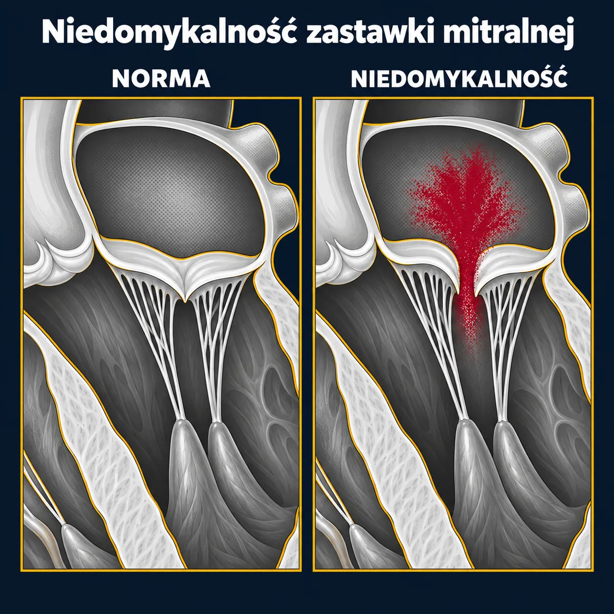
There are two fundamentally different types of this condition:
- Primary (organic) — the valve itself is damaged: most often leaflet prolapse (degenerative changes, affecting 1–2.5% of the population), a ruptured chorda tendinea (the cord-like structures holding the leaflets), and less often infective endocarditis or rheumatic disease.
- Secondary (functional) — the valve is structurally normal but does not close because the left ventricle is enlarged (after a heart attack, in cardiomyopathy) or the left atrium is enlarged (in long-standing atrial fibrillation).
Indications for a procedure in severe primary regurgitation (according to ESC/EACTS 2025):
- symptoms — breathlessness, fatigue, palpitations;
- no symptoms, but a weakened left ventricle (ejection fraction ≤ 60%) or an enlarged one (end-systolic diameter ≥ 40 mm);
- no symptoms and a normal ventricle, if atrial fibrillation, pulmonary hypertension or marked left atrial enlargement develops, and the valve is likely to allow a durable repair in an experienced centre — the 2025 guidelines give a class I recommendation here when specific criteria are met.
In secondary regurgitation, heart failure treatment is optimised first (medicines, possibly cardiac resynchronisation therapy); a procedure is considered when symptoms persist despite treatment and a benefit after the procedure is realistic.
The decision is made by the Heart Team, including a cardiologist, an interventional cardiologist, a cardiac surgeon and a heart failure specialist.
What it involves
- Valve repair (valvuloplasty) — the method of choice in primary regurgitation. It involves removing or stitching the prolapsing part of the leaflet, implanting artificial chordae (PTFE sutures) and sewing in a stabilising ring. In experienced centres more than 90% of degenerative valves are repaired; repair gives better long-term survival than replacement and does not require long-term anticoagulation.
- Valve replacement — when repair is not possible (advanced calcification, rheumatic disease, destruction after endocarditis). A biological or mechanical prosthesis is used; the subvalvular apparatus is preserved to protect left ventricular function.
- Minimally invasive access — through a right lateral mini-thoracotomy (a 5–7 cm incision between the ribs), with cannulation of the groin vessels and video assistance; the breastbone remains intact. In selected centres, robotic access. Not every patient is eligible (for example with pleural adhesions, severe atherosclerosis of the femoral arteries, or a need for simultaneous bypass grafting).
- Transcatheter edge-to-edge repair (TEER, e.g. MitraClip) — a clip that joins the two leaflets at the site of the leak is introduced through the femoral vein. It is used in patients with high surgical risk in primary regurgitation and — with suitable anatomy — in secondary regurgitation that does not respond to medicines, where it has been shown to improve survival and reduce hospital admissions.
During mitral valve surgery, atrial fibrillation ablation and left atrial appendage closure are often performed at the same time, and, if there is significant tricuspid regurgitation, tricuspid repair as well (separate pages).
What happens
Admission the day before surgery. Required tests: transoesophageal echocardiography (detailed assessment of the mechanism of the leak), coronary angiography or coronary CT angiography and, for minimally invasive access, a CT angiogram of the aorta and iliac arteries. The operation is performed under general anaesthesia with cardiopulmonary bypass and takes 3–5 hours; the result of the repair is checked during the operation with transoesophageal echocardiography. One day in intensive care.
Typical hospital stay: 5–7 days (surgery), 2–4 days (TEER).
Recovery
- After sternotomy: protect the breastbone for 6–8 weeks. After mini-thoracotomy: pain between the ribs for 2–4 weeks, but without the "breastbone" restrictions; return to activity is faster.
- After repair: anticoagulation usually for 3 months (until the ring heals in), then depending on heart rhythm; after a mechanical prosthesis — lifelong; after TEER — usually antiplatelet or anticoagulant treatment for a few months, as advised by your doctor.
- Follow-up echocardiography at 6–12 weeks, at one year, then regularly.
- Cardiac rehabilitation is recommended. Prevention of endocarditis before dental procedures.
Risks and alternatives
Approximate figures for planned valve repair in primary regurgitation: death 1–2%, stroke 1–2%, bleeding requiring re-operation 2–4%, pacemaker 2–4%, atrial fibrillation after surgery 30–40%. The risk of replacement is somewhat higher than that of repair, and surgery for secondary regurgitation carries a higher risk because of the coexisting heart failure. Durability of repair: more than 90% of patients free from re-operation at 10 years for posterior leaflet prolapse; slightly fewer for anterior or both-leaflet prolapse.
TEER: 30-day mortality about 1–2%, but some patients are left with moderate regurgitation; effectiveness depends on anatomy.
Alternatives: conservative treatment — in primary regurgitation it does not stop damage to the ventricle; in secondary regurgitation it is the foundation before any procedure; transcatheter mitral valve replacement (TMVR) — an evolving method, available in selected centres and situations.
More on how the choice between surgery and a transcatheter procedure is made →
Questions
Repair or replacement?
In primary regurgitation — repair, whenever possible. It gives better survival, preserves ventricular function and does not require long-term anticoagulation.
Is minimally invasive surgery just as effective?
Repair success rates and durability are comparable in experienced centres; the benefit is less trauma and a faster return to activity. Eligibility depends on anatomy and other conditions.
When MitraClip instead of surgery?
When surgical risk is high, or in secondary regurgitation with heart failure, provided the leaflet anatomy is suitable. In a younger patient with primary regurgitation and low risk, surgery gives a more durable result.
Can the leak come back?
After repair — in a small proportion of patients, which is why echocardiographic follow-up is a permanent part of care.
What about atrial fibrillation?
If present, ablation is usually performed during the operation and the left atrial appendage is closed — this reduces the risk of stroke and of the arrhythmia returning.