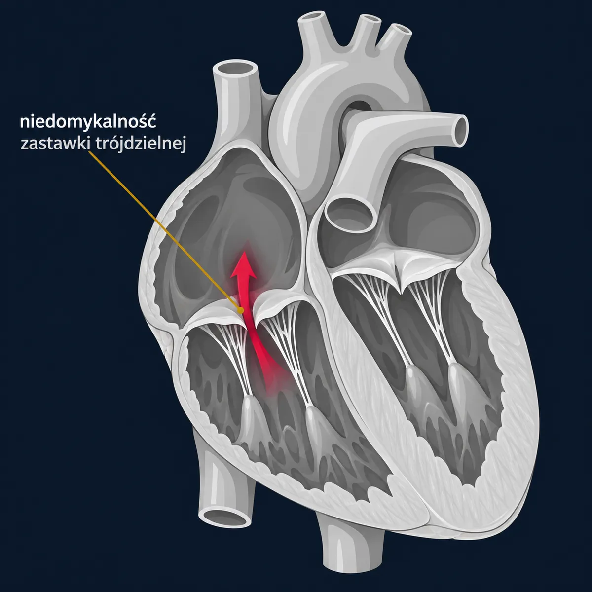
Who it is for
- Secondary (functional) regurgitation — more than 90% of cases. The valve is structurally normal, but the annulus and the right ventricle are stretched because of left-sided heart valve disease, pulmonary hypertension or long-standing atrial fibrillation (so-called atrial regurgitation).
- Primary — damage to the valve itself: infective endocarditis (common in people who inject drugs), damage by a pacemaker or defibrillator lead, injuries, congenital defects (Ebstein's anomaly), and rarely inflammatory diseases.
Indications (ESC/EACTS 2025):
- During mitral or aortic valve surgery: severe tricuspid regurgitation — always; moderate or mild, if the annulus is dilated (≥ 40 mm or > 21 mm/m² on echocardiography) or there have been symptoms of right heart failure — also recommended; adding a repair increases the risk only slightly and prevents later progression.
- Isolated severe regurgitation: in patients with symptoms (swelling, an enlarged liver, fluid in the abdomen, fatigue), before irreversible damage to the right ventricle, liver and kidneys occurs; in patients without symptoms — if the right ventricle is progressively enlarging or weakening.
- Late regurgitation after left-sided heart surgery: a transcatheter procedure or re-operation in an experienced centre is considered.
Timing is crucial in this condition: surgery in a patient with advanced right ventricular failure and a damaged liver carries a high risk.
What it involves
- Annuloplasty (ring repair) — sewing in a rigid or semi-rigid ring that reduces and stabilises the valve opening; the method of choice in secondary regurgitation. The ring leaves a gap near the atrioventricular node so as not to damage the heart's conduction system.
- Leaflet repair — for primary damage: stitching, artificial chordae, enlarging a leaflet with a patch of pericardium.
- Valve replacement — when repair is not possible; usually a biological prosthesis (on the right side of the heart, where pressure is low, mechanical prostheses are more prone to clotting).
- Transcatheter procedures — edge-to-edge repair (T-TEER, clip) and transcatheter tricuspid valve replacement (TTVR) — in patients at high surgical risk; studies in recent years have shown improvement in symptoms and quality of life, and survival data are being collected.
Tricuspid surgery is performed through a sternotomy or a right mini-thoracotomy; an isolated repair can be performed on the beating heart, without stopping the coronary circulation.
What happens
Diagnostic tests: transthoracic and transoesophageal echocardiography (including assessment of the size and function of the right ventricle), often cardiac MRI, right heart catheterisation (pressures in the lungs), assessment of the liver and kidneys. The operation is performed under general anaesthesia with cardiopulmonary bypass: 2–3 hours as an isolated procedure, longer as part of a combined operation. One day in intensive care.
Typical hospital stay: 6–8 days; with advanced right heart failure the stay may be longer, because fluid needs to be removed and liver and kidney function stabilised.
Recovery
- Restrictions after sternotomy for 6–8 weeks; shorter after mini-thoracotomy.
- Diuretics often continue for weeks or months; the dose is set by your doctor based on swelling and body weight.
- Anticoagulation: after a ring — usually 3 months (longer with atrial fibrillation); after a biological prosthesis — 3–6 months; as advised by your doctor.
- Follow-up echocardiography at 3 months and at one year, then regularly; not only the valve but also the right ventricle is assessed.
- Cardiac rehabilitation is recommended.
Risks and alternatives
Adding a tricuspid repair to mitral valve surgery increases the risk of needing a pacemaker by a few percent, but does not significantly increase mortality. Isolated tricuspid surgery has a mortality risk of 3–8% in registries — largely because patients are operated on too late, with right ventricular failure; in patients operated on early the risk is clearly lower. Main complications: atrioventricular block requiring a pacemaker (5–10%), bleeding, right heart failure, recurrent regurgitation (more often after repair without a ring).
Alternatives: conservative treatment (diuretics) — relieves symptoms but does not change the course of the disease; transcatheter procedures — in patients who cannot undergo surgery; treating the cause (e.g. atrial fibrillation ablation in atrial regurgitation, removal of a lead).
More on how the choice between surgery and a transcatheter procedure is made →
Questions
Does tricuspid regurgitation resolve on its own after mitral valve surgery?
Sometimes, yes, but in a large proportion of patients it persists or worsens, which is why the guidelines recommend a simultaneous repair when the annulus is dilated.
A pacemaker lead is damaging the valve — what then?
During surgery the lead can be repositioned or replaced with an epicardial lead (placed on the outer surface of the heart); the decision depends on the type of device.
Is the clip procedure on the tricuspid valve effective?
In well-selected patients it reduces the leak and symptoms; it cannot be performed in every anatomy (a large gap between the leaflets, a lead in the way).
Why is the liver assessed in this condition?
Because venous congestion damages the liver (so-called congestive liver), and the degree of this damage determines the surgical risk and whether it is already too late.