Who is it for
A PFO is not closed simply because it is there, but when it has probably caused a stroke. Indications (according to European guidelines and the CLOSE, RESPECT and REDUCE trials):
- a patient aged 18–60 after an ischaemic stroke or embolism of unexplained cause (cryptogenic stroke), following full investigation that has ruled out other causes (atrial fibrillation, carotid atherosclerosis, small-vessel disease);
- a PFO with "high-risk" features: a large shunt on contrast echocardiography, an atrial septal aneurysm; the RoPE and PASCAL scores are helpful, as they estimate how likely it is that the PFO caused the stroke;
- the decision is made jointly by a neurologist and a cardiologist.
A PFO is not closed routinely because of migraine, or as "prevention" in people who have not had a stroke — the evidence is insufficient. Rare indications: platypnoea-orthodeoxia syndrome, decompression sickness in professional divers.
An ASD is closed when the shunt is significant: enlargement of the right ventricle and atrium on echocardiography, a ratio of pulmonary to systemic blood flow (Qp/Qs) ≥ 1.5, symptoms (breathlessness, heart rhythm disturbances, reduced exercise capacity), provided the pressure in the lungs is not too high. The most common type — secundum ASD (ASD II), about 70% of defects — is usually suitable for catheter-based closure if it has adequate edges (rims) to anchor the occluder. Sinus venosus and primum defects, as well as very large defects or those without rims, require surgery.
What it involves
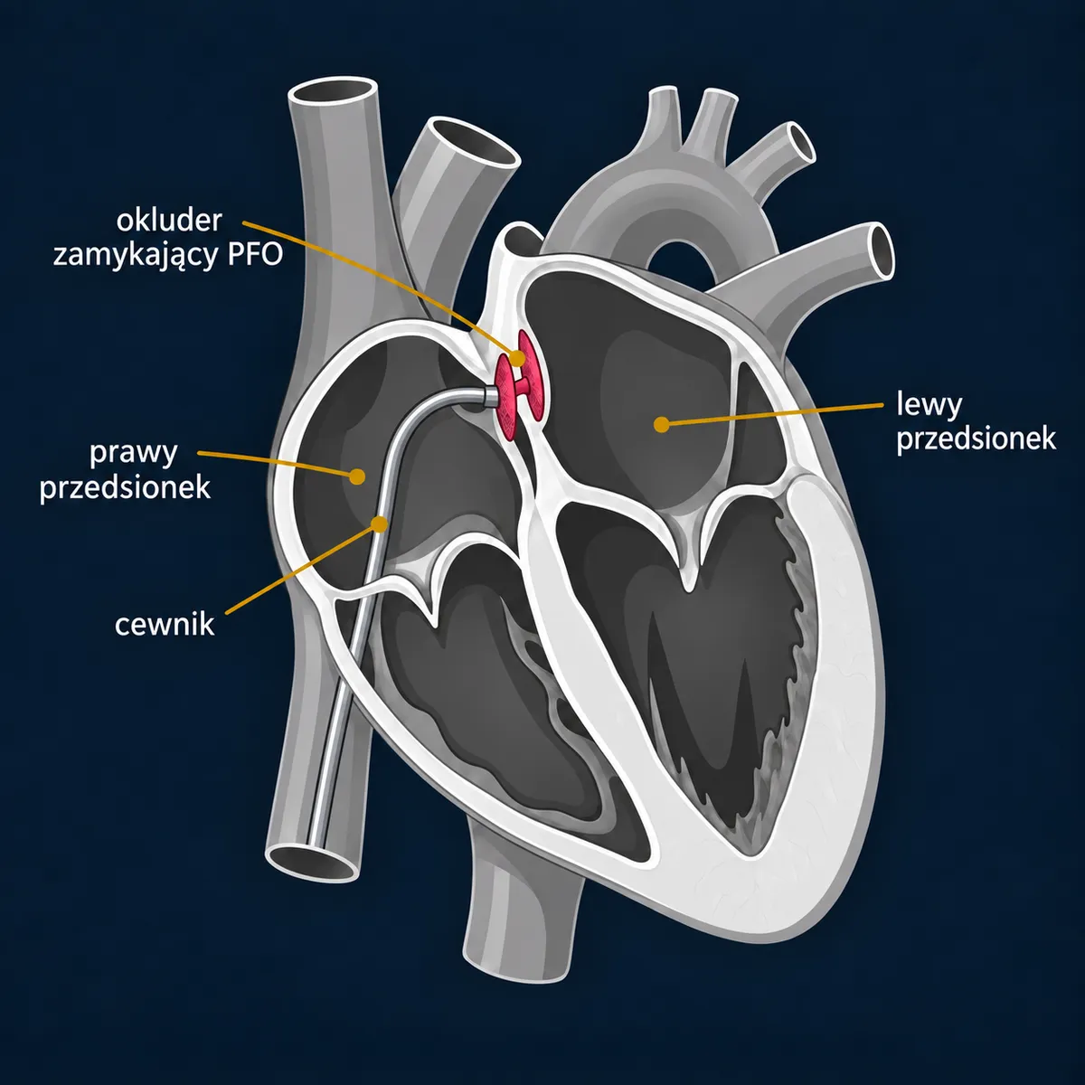
- Catheter-based closure — an occluder (two discs joined by a waist) is passed through the femoral vein and opened on both sides of the septum; over time it becomes covered with the body's own tissue. The procedure is guided by transoesophageal or intracardiac echocardiography. Local anaesthesia with sedation, or general anaesthesia.
- Surgery — through a sternotomy or a right mini-thoracotomy, using cardiopulmonary bypass: the defect is stitched closed directly or closed with a patch made from your own pericardium; in a sinus venosus defect, abnormally draining pulmonary veins are also redirected. Surgery is a definitive treatment, with very low risk and a practically zero rate of residual shunt.
What to expect
Assessment: transthoracic echocardiography with contrast (bubble test), transoesophageal echocardiography (size of the defect, rims, pulmonary veins); in patients with ASD often cardiac MRI or CT angiography and right heart catheterisation. After a stroke — full neurological work-up, including long-term heart rhythm monitoring to look for atrial fibrillation.
Catheter-based procedure: 30–60 minutes, hospital stay 1–2 days. Surgery: 2–3 hours, one day in intensive care, hospital stay 5–7 days.
Recovery
- After the catheter-based procedure: limit physical exertion for about 1–2 weeks (the groin and the septum are healing), antiplatelet treatment (usually aspirin, initially with clopidogrel) for 3–6 months until the occluder is covered with tissue, prevention of endocarditis for 6 months; echocardiography at 1, 6 and 12 months. MRI is possible (occluders are MR-conditional — according to the implant documentation).
- After surgery: protect the breastbone for 6–8 weeks (after a sternotomy); anticoagulation is usually not needed, aspirin for a few months as advised by your doctor.
- After ASD closure the right ventricle usually becomes smaller over the following months; an improvement in exercise capacity is noticeable even in patients who "had no symptoms".
Risks and alternatives
Catheter-based procedure: serious complications < 1–2% — tamponade, air embolism, displacement of the occluder (requiring removal, sometimes surgical), erosion of the atrial wall (very rare, < 0.1%, but dangerous). Temporary heart rhythm disturbances (atrial fibrillation) in the first weeks — a few per cent, more often in older patients. Any residual shunt is usually small and of no significance.
Surgery: death < 1% in young adults, atrial fibrillation after surgery, fluid around the heart (post-pericardiotomy syndrome) — a few per cent.
Alternatives for PFO after a stroke: antiplatelet treatment — trials have shown that closure reduces the risk of another stroke compared with medication alone in well-selected patients (an absolute risk reduction of about 1% per year). For ASD there is no alternative other than observation of small, insignificant defects.
More on how the choice between surgery and a transcatheter procedure is made →
Questions
I have a PFO and migraines — will closure help?
Study results are inconclusive; this is not a recognised indication. It is considered only exceptionally, in specialist referral centres.
Should a PFO be closed "just in case"?
No. One in four people has one, and in the vast majority it never causes any symptoms.
Can I play sport after closure?
Yes, after the healing period; professional diving requires additional assessment.
An ASD was found in an adult over 50 — is closure worthwhile?
Yes, if the shunt is significant and the pressure in the lungs is not too high; the benefit of less breathlessness and a lower risk of arrhythmia also applies to older patients, although atrial fibrillation present before the procedure may recur.
Is the occluder visible, and does it need replacing?
It stays in place permanently, cannot be seen from the outside and, once covered with tissue, does not need replacing.